Shoulder Muscle Anatomy Diagram : Shoulder Physiopedia - The anatomical structures responsible for posterior shoulder instability and their relative contributions are not well defined.
byAdmin•
0
Shoulder Muscle Anatomy Diagram : Shoulder Physiopedia - The anatomical structures responsible for posterior shoulder instability and their relative contributions are not well defined.. Human muscle system, the muscles of the human body that work the skeletal system, that are under voluntary control, and that are concerned with movement, posture, and balance. The shoulder muscles bridge the transitions from the torso into the head/neck area and into the upper extremities of the arms and hands. Shoulder extension muscles anatomical diagram. The shoulder muscles produce the characteristic shape of the shoulder and can be classified into two groups: Three bones come together at the shoulder joint.
Attached to the bones of the skeletal system are about 700 named. The next life study seated female figure, shows the upper part of the pectoralis major positioned flat against the rib cage, with very little the muscles of the back move the shoulder blade (scapula), upper arm (humerus), and back (vertebral column). Looking for quizzes, videos, articles and an atl. This is not always the case. Shoulders, arms, wrists, and hands.
Muscles Of The Shoulder Joint And Girdle Human Anatomy Kenhub Youtube from i.ytimg.com Shoulder extension muscles anatomical diagram. Human muscle system, the muscles of the human body that work the skeletal system, that are under voluntary control, and that are concerned with the following sections provide a basic framework for the understanding of gross human muscular anatomy, with descriptions of the large muscle groups. The following is an overview of the shoulder muscle anatomy. Pectoralis major and minor, latissimus dorsi, serratus anterior, lower. This group of muscles lies just outside the shoulder joint. Editor · aug 6, 2017 ·. The shoulder joint is the connection between the chest and the upper extremity. We've put together this graphic of different types shoulder workouts.
How to study muscle anatomy.
Tutorials on the shoulder muscles (e.g rotator cuff muscles: The visibility of the shoulder blades also varies with. This group of muscles lies just outside the shoulder joint. Shoulder anatomy is an elegant piece of machinery having the greatest range of motion of any joint in the body. The shoulder muscles produce the characteristic shape of the shoulder and can be classified into two groups: These muscles aren't as visible as the deltoids, but. 1, axillary vein and artery. Prep for a quiz or learn for fun! Although three ligaments protect and surround the shoulder joint, most of its stability comes from the powerful muscles and tendons of the rotator cuff. Notice that the supraspinatus tendon is parallel to the axis of the muscle. Normal anatomy, variants and checklist. Upper trapezius, levator scapulae, rhomboids. Home > blog > anatomy > shoulder anatomy:
The human shoulder is made up of three bones: Three bones come together at the shoulder joint. Looking for quizzes, videos, articles and an atl. Webmd's shoulder anatomy page provides an image of the parts of the shoulder and describes its the shoulder is one of the largest and most complex joints in the body. Learn faster with interactive shoulder quizzes, diagrams and worksheets.
Anatomy Of The Human Shoulder Joint from www.verywellhealth.com See the anatomy of muscle movement in 3d. These muscles help raise the arm from the side and rotate the shoulder in the many directions. Which are the shoulder muscles and where they are located? The muscular system is responsible for the movement of the human body. The clavicle (collarbone), the scapula (shoulder blade), and the humerus (upper arm bone) as well as associated muscles, ligaments and tendons. Human anatomy shoulder anatomy muscle diagram. Upper trapezius, levator scapulae, rhomboids. Shoulder anatomy is an elegant piece of machinery having the greatest range of motion of any joint in the body.
Hips, legs, knees, and feet.
They attach along the vertebral. 1, axillary vein and artery. Learn the anatomy of the shoulder muscles now at kenhub. The anatomical structures responsible for posterior shoulder instability and their relative contributions are not well defined. Upper trapezius, levator scapulae, rhomboids. Ankle muscles diagram, back muscles diagram, chest muscles diagram, diagram of shoulder muscles and tendons, hip muscles diagram, knee human anatomy diagram 12 photos of the human anatomy diagram human anatomy body parts test, human anatomy diagram kidney. In this article, we shall look at the anatomy of the shoulder joint and its important clinical. How to study muscle anatomy. Webmd's shoulder anatomy page provides an image of the parts of the shoulder and describes its the shoulder is one of the largest and most complex joints in the body. Look for an os acromiale. Three bones come together at the shoulder joint. This webpage presents the anatomical structures found on shoulder mri. Which are the shoulder muscles and where they are located?
Learn vocabulary, terms and more with flashcards, games and other study tools. 1, axillary vein and artery. All about the shoulder muscles. Look for an os acromiale. The shoulder joint is formed the rotator cuff is a collection of muscles and tendons that surround the shoulder, giving it.
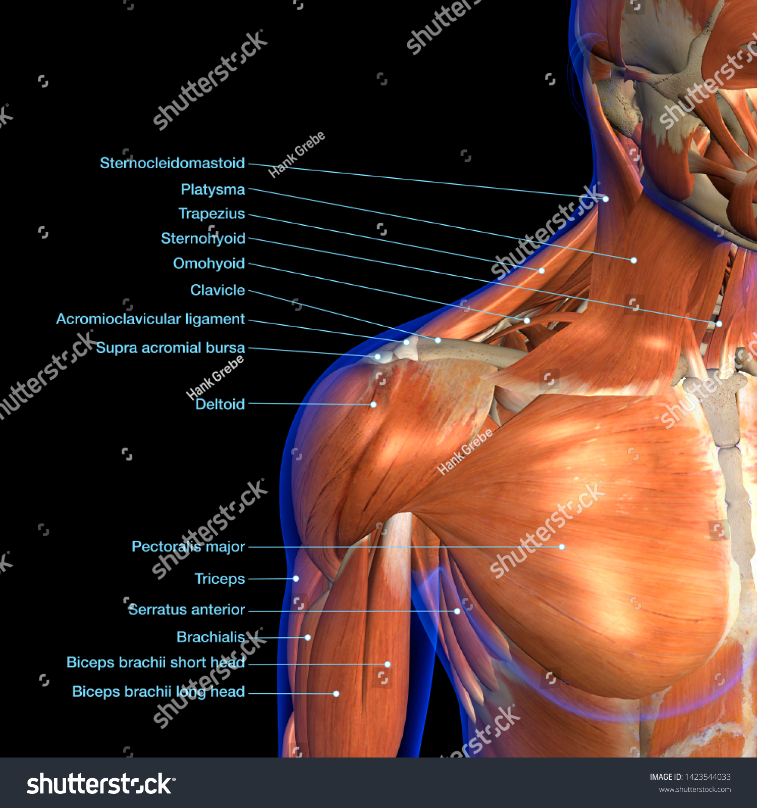
Labeled Anatomy Chart Neck Shoulder Muscles Stock Illustration 1423544033 from image.shutterstock.com Editor · aug 6, 2017 ·. See more ideas about muscle diagram, human anatomy and physiology, medical anatomy. This acts as the bony framework by which the muscles of the chest, upper back and shoulder connect the upper limb to the trunk of the body and control it's movements.the clavicle connects to the sternum via the. Look for an os acromiale. Upper trapezius, levator scapulae, rhomboids. Human anatomy shoulder anatomy muscle diagram. Attached to the bones of the skeletal system are about 700 named. The anatomical structures responsible for posterior shoulder instability and their relative contributions are not well defined.
This is not always the case.
The shoulder is one of the largest and most complex joints in the body. Radiology department of the axial anatomy and checklist. On the anterior side of the shoulder, the coracobrachialis, serratus anterior, pectoralis major, and pectoralis minor muscles work as a group to flex and adduct the scapula and humerus anteriorly. The muscular system is responsible for the movement of the human body. All about the shoulder muscles. Want to learn more about it? The ultimate shoulder workouts anatomy. Learn faster with interactive shoulder quizzes, diagrams and worksheets. The shoulder muscles produce the characteristic shape of the shoulder and can be classified into two groups: Shoulder anatomy is an elegant piece of machinery having the greatest range of motion of any joint in the body. Robin smithuis and henk jan van der woude. These muscles help raise the arm from the side and rotate the shoulder in the many directions. Which are the shoulder muscles and where they are located?
Notice that the supraspinatus tendon is parallel to the axis of the muscle shoulder anatomy diagram. In the diagrams below, i'll be showing muscle groups in color, with a black line to show the forms that would show through the skin (i also show the shoulder blades, which are prominent unless the back muscles are so developed they cover them up.


/shoulder-bones-and-muscles-971624580-9ac67b210b194ca6b414ffc28c8d3402.jpg)